


 日志正文
|
|||||
 问题:去年因为我的男友患脑干胶质瘤,最终不幸离开了。我想了解一些关于脑干胶质瘤方面的知识?谢谢! 问题:去年因为我的男友患脑干胶质瘤,最终不幸离开了。我想了解一些关于脑干胶质瘤方面的知识?谢谢!答复: 您好!脑干位于颅后窝内,俯卧于颅底蝶鞍斜坡上,上接间脑,下端在枕骨大孔处延续为脊髓,脑干向下向上分为延髓、脑桥和中脑三部分。脑干为第Ⅲ~第Ⅻ颅神经的起源地,与嗅神经及视神经有传导反射联系,是上行及下行的长束必经之处。脑干的网状结构中有呼吸及循环中枢,其网状激动系统与意识有密切关系。脑干症状复杂多样,其特点是:病变同侧的周围性颅神经麻痹和对侧的中枢性偏瘫及偏身感觉障碍—交叉性麻痹。 脑桥症状(1)颅神经症状:脑桥病变引起的三叉神经症状以病灶侧面部感觉障碍为主,角膜反射减低或丧失,同侧咀嚼肌萎缩且肌力弱,张口下颌偏向患侧,外展神经麻痹,眼球内斜。(2)感觉障碍:感觉障碍程度不一,有的完全缺失,有的轻度减退。股体感觉障碍及面部感觉可以呈交叉状态。肢体感觉症状又表现为分离性感觉障碍。(3)运动麻痹:多于病灶对侧出现偏瘫。脑桥下部病变时,病侧出现面神经麻痹,对侧出现偏瘫,同时病灶侧尚有外展神经麻痹。(4)小脑症状:小脑症状为为脑桥病变重要的症状之一 ,脑桥与小脑关系密切,脑桥病变时病灶侧出现共济失调及其他小脑症状。(5)精神及睡眠障碍:脑桥病变因损伤脑干网状结构可出现精神障碍、智力下降及睡眠障碍,起初淡漠、嗜睡、悲痛易哭、继之则好动,语言讷吃。 治疗由于目前的显微神经外科技术比较发达,能够进行一部分病人的全切手术。 同时采用节拍式化疗和血管抑制治疗。是可以对一部分患者进行根治的。 下面是一些详细的脑干区域的解剖结构和功能: 
The figure below on the right shows the entire ventral surface of the brain stem. The lines and arrows connecting the ventral view with the three MRI images are to help you understand what is left and right. Do not worry about the bumps, grooves and nerves shown on this ventral view, that will come later. Right now just orient yourself!! Both the dorsal and ventral surfaces of the brain stem are shown below. We already know what is right and left on the ventral view. Now, take the page out of your notebook and fold it along the dotted line (yes, this is medical school!) and you can see that what lies beneath (is dorsal) the right side of the ventral aspect of the brain stem is the right side of the dorsal aspect of the brain stem, and what lies below the left side of the ventral aspect of the brain stem is left side of the dorsal aspect of the brain stem. Like in the spinal cord, in an axial MRI section of the brain stem ventral is up and dorsal is down. Again we will follow tradition (all neuro textbooks!) and “flip” the section so ventral is down and dorsal up. However, make sure to keep the right/left designation of the axial section MRI. That is, the right side of the patients brain is on your left.
As mentioned earlier, the brain stem includes the (1) medulla, the (2) pons and the (3) midbrain. The brain stem contains 10 cranial nerves, and most of the motor and sensory systems pass through this important region. It is a relatively small region (approximately 7 cm long) that links the forebrain (i.e., cerebral cortex) and the spinal cord and all messages going between the two areas must go through the brain stem.
例一:脑干海绵状血管瘤切除
  
例二:脑干斜坡部脊素瘤   脊索瘤是起源于胚胎脊索结构的残余组织的先天性良性肿瘤,是一种破坏性肿瘤,发病率低,有报告占脑瘤的0.15-0.2%,发病年龄高峰为30-40岁,平均年龄为35-40岁,男性比女性多见。颅内脊索瘤多起自斜坡中线部位,位于硬膜外,缓慢浸润性生长。位于蝶枕部占35%,脊柱部占15%,骶尾部最多,占50%。病程长,平均在3年以上,头痛为最常见症状,头痛性质是持续性钝痛,常为全头痛,也可向枕部或颈部扩展,因肿瘤部位,肿瘤的发展方向不同其临床表现各有所不同。斜坡部脊素瘤:主要表现为脑干受压症状,即步行障碍,锥体束征,外展、面神经功能损害。由于肿瘤发生于颅底,可引起交通性脑积水,如:肿瘤向桥小脑角发展,出现听觉障碍,耳鸣、眩晕,若起源于鼻咽壁远处,常突到鼻咽引起鼻不通气、疼痛,可见脓性或血性分泌物。治疗:一般采用外科手术和放射治疗,但现在的治疗结果是令人失望的,颅底肿瘤的主要肿块,靠近脑干的肿瘤均不易暴露,放疗也不敏感,预后不良。中医治疗:抗瘤正脑系列的配伍应用,适用于未行手术或手术部分切除,术后复发,X-刀、γ-刀,放化疗后患者用药3-6个月可消除症状,使瘤体缩小或消失,预防复发,临床应用多年来疗效确切。 脊索瘤是起源于胚胎脊索结构的残余组织的先天性良性肿瘤,是一种破坏性肿瘤,发病率低,有报告占脑瘤的0.15-0.2%,发病年龄高峰为30-40岁,平均年龄为35-40岁,男性比女性多见。颅内脊索瘤多起自斜坡中线部位,位于硬膜外,缓慢浸润性生长。位于蝶枕部占35%,脊柱部占15%,骶尾部最多,占50%。病程长,平均在3年以上,头痛为最常见症状,头痛性质是持续性钝痛,常为全头痛,也可向枕部或颈部扩展,因肿瘤部位,肿瘤的发展方向不同其临床表现各有所不同。斜坡部脊素瘤:主要表现为脑干受压症状,即步行障碍,锥体束征,外展、面神经功能损害。由于肿瘤发生于颅底,可引起交通性脑积水,如:肿瘤向桥小脑角发展,出现听觉障碍,耳鸣、眩晕,若起源于鼻咽壁远处,常突到鼻咽引起鼻不通气、疼痛,可见脓性或血性分泌物。治疗:一般采用外科手术和放射治疗,但现在的治疗结果是令人失望的,颅底肿瘤的主要肿块,靠近脑干的肿瘤均不易暴露,放疗也不敏感,预后不良。中医治疗:抗瘤正脑系列的配伍应用,适用于未行手术或手术部分切除,术后复发,X-刀、γ-刀,放化疗后患者用药3-6个月可消除症状,使瘤体缩小或消失,预防复发,临床应用多年来疗效确切。   例三:脑干肿瘤 18岁的贵州女孩芳芳(化名)却因脑干肿瘤不得不终止学业。3月8日,成都军区总医院成功地为芳芳进行了脑干肿瘤切除手术。不久以后,她就将重返课堂。
医生仔细检查病情
女孩脑干肿瘤的位置 初期症状为右腿行动不便,一只眼睛眼球无法移动,在北京的一家医院查出了其脑干部位肿瘤。因为这个脑干肿瘤切除手术的危险系数高
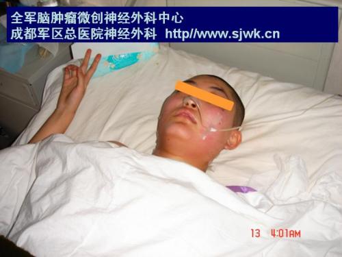
手术后躺在病床上的女孩 3月8日上午10点,成都军区总医院总共花了3个多小时实施该手术。手术中,运用了电子显微镜、导航系统等高科技仪器,确保了手术的成功。5天过去了,芳芳已经坐起来,能够自己进一些流食。再过几天就可以下地活动了。
最后修改于 2008-07-01 06:46
阅读(?)评论(0)
|
|||||


 Details regarding the organization of the brain stem will be presented by discussing the nuclear groups and fiber tracts that are present at 10 different caudal to rostral levels.


评论 想第一时间抢沙发么?